1. Why diagnosis is more than “it’s just stress” or “it’s genetic”
Most people with hair loss have heard at least one of these lines:
- “It’s just stress.”
- “It’s hormones.”
- “It’s in your genes, nothing to do.”
Each of these may contain a fragment of truth; however, none of them is a diagnosis.
In reality, hair loss is an umbrella term. Underneath it are multiple distinct conditions, some reversible, some not, some requiring urgent treatment, others more tolerant of watchful waiting. The purpose of a proper assessment is to:
- identify which process or processes are present,
- quantify severity and pattern,
- look for underlying contributors, and
- separate self-limiting states from those needing longer-term management.
It is less about guessing the “cause” in a sentence, and more about building a structured picture.
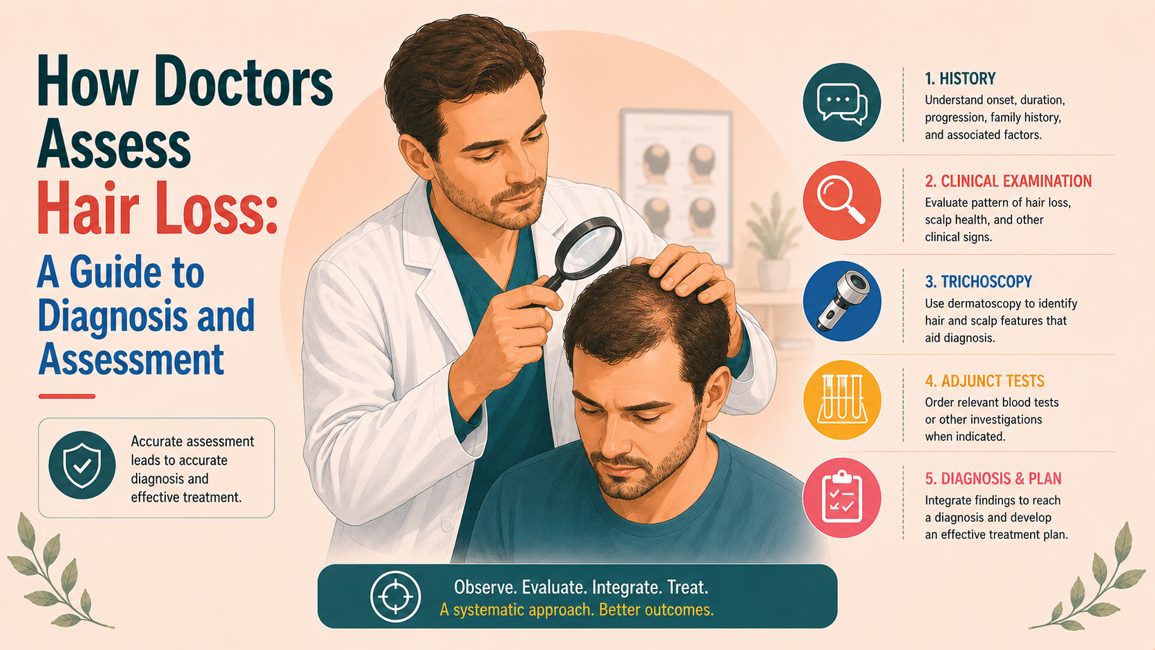
2. The starting point: a structured history
The first diagnostic tool is conversation. A good history will narrow down the diagnosis significantly.
2.1 Onset and timing
Your doctor will want to know:
- When did you first notice something was different?
- Did it start suddenly or gradually?
- Has it been stable, slowly progressive, or did it accelerate at a particular time?
Sudden shedding a few months after a major illness (e.g. telogen effluvium) points in a very different direction to gradual frontal thinning since your early twenties (likely male pattern hair loss).
2.2 Pattern as you perceive it
What you may have noticed matters:
- more hair on the pillow or in the drain,
- widening of the parting,
- receding hairline,
- specific patches in the scalp, beard or brows,
- change in ponytail thickness.
Patient language often provides helpful clues about whether we are dealing with shedding, pattern loss, patchy loss, or a combination of these.
2.3 Triggers and context
A clinician will explore:
- recent illnesses, surgery, high fevers or infections,
- pregnancies, miscarriages, terminations, breastfeeding,
- recent weight changes or dieting patterns,
- psychological stressors, bereavements, major life events,
- new medications or recent changes (including contraception, retinoids, psychiatric or endocrine drugs).
Many types of hair loss are reactive; that is, they occur several weeks to months after a stressor. Matching the timing helps uncover those links.
2.4 Hair care and styling practices
This includes:
- typical hairstyles (tight ponytails, braids, extensions, man-buns),
- use of relaxers, bleaching, colouring, keratin treatments,
- heat styling habits.
Chronic traction or chemical overuse can create very specific patterns of loss that require different management from hormonally driven conditions.
2.5 Family history
Pattern hair loss, some autoimmune diseases and some scarring alopecias have a familial element. Knowing who in your family lost hair, when and how, helps calibrate risk and expectations.
2.6 General health, hormones and nutrition
Questions may cover:
- has lost hair, when, and howmenstrual regularity, menopausal status, libido, erectile function,
- acne, hirsutism, weight gain or loss,
- changes in energy, bowel habits, heat or cold tolerance (for thyroid),
- known iron deficiency or anaemia, heavy periods, dietary restrictions.
The aim is not to pathologise every variation, but to identify genuine hormonal or nutritional contributors that, if present, ought to be addressed.
3. Clinical examination: pattern, scalp and shafts
Once the history is clear, the scalp and hair are examined.
3.1 Global inspection
The clinician will step back and look at:
- overall distribution of hair,
- specific areas of thinning or baldness,
- symmetry (diffuse vs focal, frontal vs crown vs occipital),
- whether the hairline is intact, receding, or scarred.
In men, a Hamilton–Norwood pattern may be obvious. In women, central thinning, consistent with Ludwig or Sinclair patterns, may be evident. Patchy areas without follicles suggest something different again (likely alopecia areata).
3.2 Scalp skin
The scalp itself is assessed for:
- redness, scale, flaking or crusting,
- follicular plugging,
- pustules or exudate,
- areas of shiny atrophic skin lacking visible follicles,
- tenderness when palpated.
These features help distinguish scarring from non-scarring conditions and suggest whether infection, psoriasis, seborrhoeic dermatitis, or autoimmune disease may be involved.
3.3 Hair shafts
Individual shafts are examined for:
- breakage versus shedding from the root,
- shaft abnormalities (kinking, twisting, nodes, beading),
- changes in diameter between hairs in the same area.
This is where shaft disorders, chemical damage and mechanical trauma become evident.
3.4 Simple bedside tests
Several quick tests can be done in the clinic:
- Pull test – gentle traction on a small bundle of hairs to see how many come away. A strongly positive test suggests active shedding.
- Tug test – assessing shaft resilience by pulling from both ends.
- Card test – placing hair against a contrasting card to assess density and shaft calibre.
- Part-width assessment – visually estimating or photographing the part to track changes over time.
These are rough tools, but when interpreted alongside the history and pattern, they add useful information.
4. Trichoscopy: dermoscopy for hair and scalp
Trichoscopy is dermoscopy applied to the scalp and hair. It uses a handheld or video dermatoscope to magnify the scalp several times, often with polarised light.
Trichoscopy is covered in more detail in our separate article, linked below.
4.1 Why it matters
Trichoscopy has become an essential part of hair loss diagnosis because it allows:
- direct visualisation of follicular openings,
- assessment of shaft thickness variability,
- identification of yellow dots, black dots, peripilar signs,
- detection of early scarring changes that are not obvious to the naked eye.
Patterns of trichoscopic findings differ between:
- androgenetic alopecia,
- telogen effluvium,
- alopecia areata,
- traction, trichotillomania, tinea capitis,
- lichen planopilaris, frontal fibrosing alopecia, CCCA and other scarring conditions.
This makes trichoscopy invaluable for differentiating look-alike conditions and for catching scarring processes early, when treatment can still preserve remaining follicles.
4.2 Quantitative trichoscopy
In some clinics, trichoscopy images are analysed digitally to count hairs per cm² and measure shaft diameters. This can:
- provide objective baselines,
- help monitor responses to treatment over months,
- reduce reliance on subjective memory of “how it used to look”.
It is not essential in every case, but it is increasingly used in specialist practice.
5. Severity grading and documentation
Diagnosis is one thing; documenting severity and change over time is another.
5.1 Clinical scales
Common scales include:
- Hamilton–Norwood (for male pattern hair loss),
- Ludwig and Sinclair (for female pattern hair loss),
- SALT (Severity of Alopecia Tool) for alopecia areata.
These allow clinicians to communicate with one another and to track progression or improvement in a structured manner.
5.2 Photographic records
Standardised photographs are a cornerstone of follow-up. Ideally:
- taken under similar lighting,
- with a consistent camera angle and distance,
- showing frontal, vertex, temporal and occipital views,
- sometimes with parted views of specific areas.
These records are far more reliable than memory for detecting subtle changes.
5.3 Imaging-based measures
More specialised techniques include:
- Phototrichogram – clipping a small patch, photographing it at defined intervals and using software to assess density and anagen/telogen ratio.
- Trichogram – an older technique involving plucking hairs and examining roots microscopically to estimate growth phase ratios.
- Cross-sectional trichometry – measuring the cross-sectional area of hair bundles to estimate bulk.
These methods are more often used in research or specialist settings than in routine general practice, but the principle is the same: to quantify what is happening rather than merely describe it.
6. Blood tests: useful, sometimes; conclusive, never
Blood tests are prone to being over-ordered or over-interpreted in hair loss. Particularly for pattern hair loss, they are rarely beneficial.
6.1 When they are useful
They are most helpful when:
- there is diffuse shedding (suggestive of telogen effluvium),
- symptoms point to systemic disease (fatigue, weight change, palpitations, menstrual changes),
- clinical signs suggest nutritional or endocrine issues.
Commonly requested tests include:
- full blood count and ferritin,
- thyroid function tests,
- vitamin D and, selectively, other micronutrients,
- androgen panel (in women with suspected hyperandrogenism),
- prolactin, fasting glucose/lipids in relevant contexts.
Abnormalities here can explain or contribute to shedding and are often treatable.
6.2 What they cannot do
Blood tests cannot:
- diagnose androgenetic alopecia (there is no “baldness blood test”),
- distinguish scarring from non-scarring alopecia,
- replace a careful look at the scalp.
Normal bloods do not rule out hair loss, and abnormal bloods do not automatically explain it. They are an adjunct, not a shortcut.
7. Scalp biopsy: when and how
A scalp biopsy is a small sample of skin and hair follicles taken under local anaesthetic, usually with a 4 mm punch. It is not needed in every case.
Scalp biopsy is covered in more detail in our separate article, linked below.
7.1 Indications
Biopsy is considered when:
- the diagnosis is unclear after history, examination and trichoscopy,
- a scarring alopecia is suspected,
- there is atypical patterning or mixed features,
- treatment decisions hinge on distinguishing between similar-seeming conditions.
7.2 Technique in outline
- The biopsy is usually taken from an active edge of hair loss rather than the centre of a bald patch.
- One or two punches may be taken, sometimes processed as horizontal and vertical sections for comprehensive analysis.
- Histopathology can reveal:
- whether follicles are scarred or intact,
- the phase distribution of follicles,
- where inflammatory cells are located (around the bulb, isthmus, and infundibulum),
- patterns of fibrosis or interface change.
Biopsy is particularly valuable in separating:
- lichen planopilaris and frontal fibrosing alopecia,
- central centrifugal cicatricial alopecia,
- discoid lupus and other scarring processes,
- from non-scarring mimics.
It is a minor procedure, but one that patients rightly want explained and justified. When used thoughtfully, it can prevent years of misdirected treatment.
8. Diagnostic pathways for common scenarios
To make this less abstract, consider a few archetypal cases.
Young man with a receding hairline
- Gradual thinning at temples and vertex over years, strong family history, healthy scalp, classic pattern on examination and trichoscopy.
- Blood tests: not routinely required if there are no systemic symptoms.
- Diagnosis: androgenetic alopecia.
- Plan focuses on medical and, if desired, surgical options; assessment is straightforward.
Woman in her thirties with diffuse thinning and heavy periods
- Widening parting and excess shedding over several months, history of heavy menstrual bleeding, and tiredness.
- Exam: central thinning consistent with early FPHL plus diffuse shedding.
- Bloods: iron deficiency anaemia found.
- Diagnosis: FPHL with superimposed telogen effluvium from iron deficiency.
- Plan: treat iron deficiency, consider starting minoxidil, and address menstrual causes.
A woman in her fifties with a receding hairline and eyebrow loss
- Hairline marching backwards over a few years, eyebrows thinning or gone, scalp feels tight or itchy.
- Trichoscopy: perifollicular scale and erythema at the margin, loss of follicles in scarred band.
- Biopsy: lichen planopilaris / frontal fibrosing alopecia pattern.
- Diagnosis: frontal fibrosing alopecia.
- Plan: anti-inflammatory and immunomodulatory therapy to halt progression; transplantation only considered very cautiously, if at all.
Man with irregular patches in beard and scalp
- Sudden appearance of smooth bald spots in the beard, one in the scalp, no symptoms.
- Exam and trichoscopy: exclamation-mark hairs, black dots, no scarring.
- Diagnosis: alopecia areata (patchy).
- Plan: discuss natural history, options including topical or intralesional steroids, and, in more extensive cases, newer systemic agents.
In each case, the diagnostic pathway is tailored, but the underlying framework – history, pattern recognition, trichoscopy, selective tests, and biopsy where indicated – is the same.
9. What patients can do before an assessment
You do not have to arrive “prepared” for your scalp to be taken seriously, but a few things can make the consultation more productive:
- Note roughly when you first noticed changes and any major life events or health issues in the preceding six to twelve months.
- Bring a list (or photos) of current medications and supplements, including hormonal contraception.
- Think about family history: who in your family lost hair, at what age, and in what pattern.
- If possible, bring older photos that show your hair a few years ago.
10. Key takeaways
- “Hair loss” is not a diagnosis. A structured assessment distinguishes between multiple conditions that happen to share thinning as a symptom.
- Good diagnosis relies first on a thoughtful history and careful examination of pattern, scalp and shafts; blood tests and biopsies are useful adjuncts but are not necessary in the majority of cases.
- Trichoscopy has become a central tool in modern hair medicine, allowing non-invasive differentiation between scarring and non-scarring processes.
- Precise diagnosis matters because scarring alopecias need early, aggressive treatment to preserve follicles, whereas reactive sheds and pattern hair loss are managed very differently.
- You are entitled to an explanation that goes deeper than “it’s just stress” or “it’s in your genes”.
Once the diagnosis is clear and documented, treatment becomes a targeted conversation rather than guesswork.

![[headshot]](https://cdn.prod.website-files.com/68207da82e5b8c350c67932f/68c6a7436a552c0fe87a4da7_Screenshot%202024-11-27%20at%2011.38.11.png)