1. What a good examination is trying to achieve
When a dermatologist or GP examines someone with hair loss, they are not simply confirming that hair has gone. The purpose is to:
- determine what type of hair loss is present (for example, androgenetic alopecia, telogen effluvium, alopecia areata, scarring alopecia);
- distinguish between scarring and non-scarring processes;
- gauge severity and distribution, which informs treatment choices;
- identify signs of underlying systemic disease;
- and document a baseline against which future change can be measured.
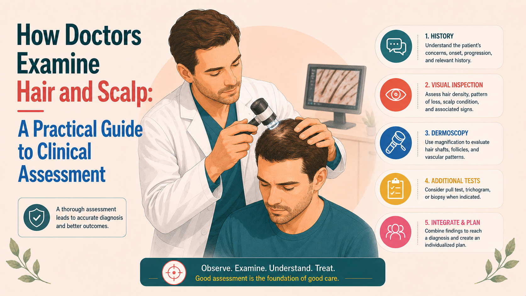
Most of this can be achieved with a structured clinical assessment, supplemented in selected cases by trichoscopy, blood tests and biopsy.
2. Global inspection: pattern, distribution and density
The first step is to stand back and simply look.
2.1 Men
In men, the examiner will look for:
- recession at the temples and frontal hairline;
- thinning at the vertex (crown);
- the relationship between frontal and vertex loss (for example, are they separate, or have they merged);
- preservation of the occipital fringe;
- overall density and how abruptly it changes from one area to another.
This allows the clinician to classify the pattern using scales such as Hamilton–Norwood (e.g. II, III, IV, V), which helps differentiate male pattern hair loss from diffuse thinning or patchy processes.
2.2 Women
In women, attention is usually directed to:
- the central parting and whether it is widening;
- whether there is frontal accentuation (“Christmas tree” pattern) or relatively uniform thinning across the mid-scalp;
- preservation of the frontal hairline and temporal areas;
- sparing of the occipital scalp.
These observations guide classification using the Ludwig or Sinclair scales (for example, Sinclair 2–5) and help distinguish female pattern hair loss from chronic telogen effluvium, diffuse alopecia areata or scarring disease.
2.3 Children and adolescents
In younger patients, pattern hair loss is less common, so clinicians often look first for:
- discrete patches of alopecia (suggestive of alopecia areata or tinea capitis);
- broken hairs with scaling and lymphadenopathy (tinea);
- unusual patterns at traction sites (tight hairstyles);
- or more generalised thinning indicative of systemic illness.
Visual distribution, therefore, provides the first layer of differential diagnosis before any tests are done.
3. Scalp skin: looking beyond the hair
A careful examination of the scalp skin is essential, because many diagnoses depend as much on what the scalp looks and feels like as on the amount of hair present.
Clinicians look for:
- Erythema (redness) – diffuse or perifollicular redness can suggest inflammatory processes such as seborrhoeic dermatitis, psoriasis, lichen planopilaris or discoid lupus.
- Scaling or flaking – fine, greasy scale suggests seborrhoeic dermatitis; thicker, adherent scale, particularly around follicles, may indicate psoriasis or scarring conditions.
- Follicular openings (ostia) – preserved in non-scarring alopecias (e.g. androgenic alopecia, telogen effluvium, alopecia areata); often reduced or absent in scarring alopecias, where skin may appear smooth and shiny.
- Perifollicular scale or “collarettes” – characteristic of some scarring conditions (lichen planopilaris, frontal fibrosing alopecia, central centrifugal cicatricial alopecia).
- Pustules or crusts – point towards infectious or neutrophilic processes (folliculitis decalvans, dissecting cellulitis, tinea capitis in children).
- Atrophy or textural change – thinning of the skin, visible veins, loss of normal skin markings suggest scarring.
- Symptoms – patients’ reports of burning, tightness or pain often accompany inflammatory or scarring disease.
This shifts the clinician from “how much hair is there?” to “what is the scalp environment like?” This is critical for determining whether aggressive anti-inflammatory treatment is needed to preserve remaining follicles.
4. Hair shaft inspection: calibre, breakage and heterogeneity
After looking globally, examiners pay attention to individual hairs.
Key features include:
- Calibre variation – in androgenetic alopecia and FPHL, miniaturisation leads to a mixture of thick terminal hairs and finer, shorter intermediate or vellus-like hairs in the same area. A visible diversity in shaft diameter is a hallmark of these conditions.
- Breakage vs shedding from the root – broken hairs of varying lengths, especially in irregular patches, point towards shaft fragility, chemical/heat damage or trichotillomania. Hairs shed with intact bulbs indicate follicular cycling changes (TE, AA).
- Shaft abnormalities – kinking, twists, nodes, “bamboo hair”, beading and other structural signs point to specific hair shaft disorders (monilethrix, trichorrhexis nodosa, pili torti).
- Length patterns – short, bristly hairs along the hairline may suggest traction; uniform short hairs following chemotherapy suggest anagen effluvium.
Clinicians may pick up small bundles of hair and examine them against a contrasting background or use a magnifying lens for more detail.
5. Simple bedside tests
A number of quick tests can be done in the consulting room to quantify or characterise shedding.
5.1 Hair pull test
The hair pull test is performed by grasping a small bundle of hairs (often 50–60) between the thumb and fingers and gently pulling along their length.
- A negative test (0–2 hairs extracted) suggests no active, excessive shedding at that site.
- A positive test (more hairs extracted) suggests increased telogen shedding (TE, AA) or fragile shafts.
The site of the pull matters: positive tests in affected areas but not in unaffected zones can help localise active disease.
5.2 Tug test
Pulling on both ends of a hair can help detect shaft fragility. If hairs break easily under mild tension, an underlying structural problem or external damage may be present.
5.3 Part-width and ponytail measurements
Although more crude, clinicians may compare the width of the central part with what is expected for the patient’s hair calibre and density, and ask about changes in ponytail circumference in women with longer hair.
These measures, particularly when tracked over time, can reveal significant trends.
6. Looking beyond the scalp
Hair loss is not always confined to the scalp. A thorough examination may include:
- Eyebrows and lashes – thinning or loss can suggest frontal fibrosing alopecia, alopecia areata, hypothyroidism or chemotherapy effects.
- Beard in men – patchy beard loss often accompanies alopecia areata; facial hair can also be a recipient or donor in transplantation decisions.
- Body hair – generalised loss may occur in alopecia universalis, endocrine disorders or certain systemic diseases.
- Nails and skin – nail pitting and ridging in alopecia areata; malar rash or discoid lesions in lupus; hyperpigmented knuckles in nutritional deficiency; papulosquamous plaques in psoriasis.
These findings can prompt diagnostic thinking beyond primary hair conditions toward systemic or autoimmune causes.
7. Context-specific nuances: men, women and children
While the basic elements of examination are similar, there are nuances by group.
7.1 Men
In adult men, the most common diagnosis is androgenetic alopecia. Examination focuses on:
- confirming the pattern and ruling out mimickers (for example, diffuse alopecia areata incognita, TE);
- assessing miniaturisation and donor status;
- looking for evidence of concomitant inflammatory or scarring conditions;
- and, where indicated, examining for signs of endocrine or metabolic disease.
Clinical appearance plus history is often sufficient for the diagnosis of AGA; atypical features guide additional tests.
7.2 Women
In women, the differential is broader and scarring alopecias are relatively more frequent.
Examination, therefore, pays particular attention to:
- distinguishing FPHL from chronic TE and diffuse AA;
- detecting subtle scarring signs at the frontal hairline or crown;
- looking for signs of androgen excess (hirsutism, acne), thyroid disease and nutritional deficits;
- and assessing traction patterns in those wearing tight styles, weaves or extensions.
Guidance from Olsen, Herskovitz, Ramos, and others emphasises that a careful clinical examination (pattern, scalp, shafts) plus selective testing often suffices to distinguish FPHL from its common mimics.
7.3 Children and adolescents
In children, tinea capitis, alopecia areata, traction alopecia and trichotillomania are prominent considerations. Examination needs to be:
- gentle, with attention to pain and cooperation;
- sensitive to psychosocial context, particularly in suspected hair-pulling;
- alert to lymphadenopathy, kerion formation or other signs of infection.
Here, examination often dictates whether to treat immediately (e.g. systemic antifungals for tinea), investigate further, or refer promptly.
8. Documentation: why it matters
A good examination does not end with observation; it is recorded in a way that supports follow-up.
Clinicians may:
- describe density, pattern and scalp features in words;
- assign severity grades (Norwood–Hamilton, Ludwig, Sinclair, SALT);
- take standardised photographs for future comparison;
- and, increasingly, capture dermoscopic images.
The purpose is to create a baseline. At future visits, this allows both doctor and patient to see whether patterns have evolved, shedding has reduced, or treatments are stabilising or improving the situation.
Patients often underestimate positive change and overestimate deterioration, particularly when anxious. Objective documentation anchors the discussion.
9. How examination feeds into further tests
A structured clinical assessment guides next steps:
- In classic AGA/FPHL without red flags, examination plus history and physical examination may be sufficient to initiate therapy without extensive investigations.
- In diffuse shedding, examination helps decide whether to look for TE triggers and nutritional or endocrine contributors.
- In suspected scarring alopecia, specific clinical signs (perifollicular scale, loss of follicular openings, atrophy) prompt early trichoscopy and often scalp biopsy.
- In alopecia areata, characteristic patches plus examination of the nails and other hair-bearing areas can avoid unnecessary blood tests unless systemic disease is suspected.
Guidelines underpin this approach: history and examination first, investigations directed by identified patterns rather than blanket testing.
10. For patients: what to look for in your own consultation
While you cannot examine your own scalp the way a dermatologist can, recognising the elements of a thorough exam can help you judge the quality of the care you receive.
It is reasonable to expect that your clinician will:
- look at your entire scalp, not only the area you first point out;
- inspect the scalp skin as well as the hair;
- check, at least briefly, your brows, lashes and nails;
- perform a simple pull test if shedding is a key concern;
- discuss what they see in terms you can understand – pattern, density, signs of inflammation or scarring.
If you leave a consultation with only a quick glance at the crown and no explanation beyond “you’re just thinning”, it is appropriate to seek a more detailed assessment or a second opinion, particularly if you are concerned about scarring or unusual patterns.

![[headshot]](https://cdn.prod.website-files.com/68207da82e5b8c350c67932f/68c6a7436a552c0fe87a4da7_Screenshot%202024-11-27%20at%2011.38.11.png)