1. What a scalp biopsy is
A scalp biopsy is a small sample of skin, usually 4 mm in diameter, taken under local anaesthetic. It includes the full thickness of the scalp skin, the hair follicles, and a portion of the underlying subcutaneous fat.
The sample is processed and examined by a dermatopathologist. The aim is not simply “to see if there is hair loss”. It is to:
- distinguish scarring from non-scarring alopecia;
- identify the type and location of inflammation, if present;
- assess follicle numbers, size and growth phase distribution;
- and narrow down the diagnosis when the clinical picture alone is not definitive.
In many cases (for example, classic male pattern hair loss or straightforward postpartum shedding), a biopsy is not necessary. But in more complex or atypical presentations, biopsy can prevent years of uncertainty or misdirected treatment.
2. When is a scalp biopsy indicated?
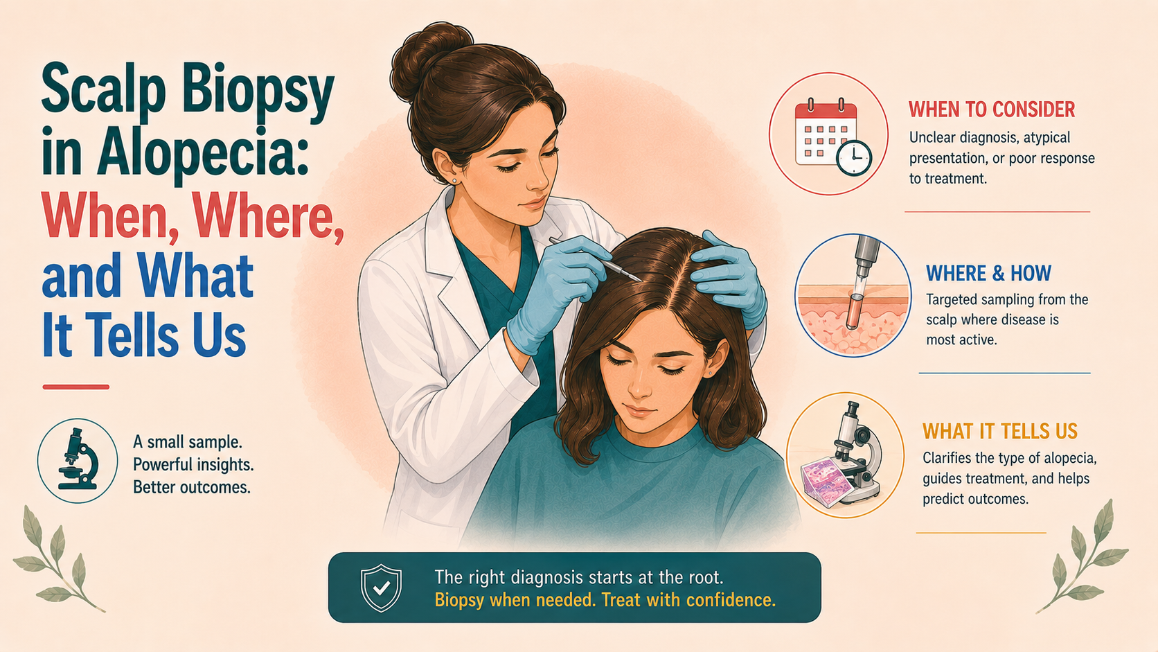
A biopsy is considered when the answer to “what is causing this hair loss?” remains uncertain after careful history, examination, and trichoscopy.
2.1 Suspected scarring (cicatricial) alopecia
If there are signs that follicles are being permanently destroyed, such as:
- areas of shiny, atrophic scalp with loss of follicular openings;
- perifollicular scale and erythema;
- symptoms such as burning, tightness or pain;
- or a rapidly receding hairline with textural changes (as in frontal fibrosing alopecia),
then a biopsy is usually recommended. This is because scarring alopecias (lichen planopilaris, frontal fibrosing alopecia, CCCA, discoid lupus, folliculitis decalvans, dissecting cellulitis) require early, targeted anti-inflammatory treatment. Additionally, diagnosis based on appearance alone can be challenging, and histology helps decide which immunomodulatory regimen is most appropriate.
2.2 Atypical or mixed patterns
A biopsy may also be suggested when:
- there is diffuse thinning that does not clearly fit androgenetic alopecia, telogen effluvium or alopecia areata;
- trichoscopic findings are equivocal or show overlapping features;
- patterns suggest more than one process (for example, FPHL plus suspected lichen planopilaris).
In these situations, a biopsy can confirm miniaturisation typical of androgenetic alopecia, detect superimposed inflammatory changes indicative of scarring disease, or reveal alopecia areata incognita when patches are not obvious.
2.3 Lack of response to appropriate treatment
Where an apparently straightforward diagnosis has been treated appropriately but the response is absent or the progression is faster than expected, a biopsy may be used to check whether a second process (for example, early scarring) has been missed.
2.4 Paediatrics and special cases
In children, biopsy is used more sparingly but may be considered when:
- tinea capitis, alopecia areata, trichotillomania and other causes have been evaluated;
- scarring processes are suspected;
- or diagnosis remains unclear despite non-invasive assessment.
Overall, a biopsy is not always necessary; it is a tailored decision driven by diagnostic uncertainty or concern about irreversible disease.
3. Where to biopsy: choosing the right site
Sampling error is a major reason biopsies can mislead. Choosing the correct site is therefore necessary.
3.1 Active margin vs end-stage centre
In suspected scarring alopecia, the ideal site is at the active edge of a lesion, where hair is still present, along with redness, scale, or symptoms.
Taking a biopsy from the completely bald centre of a long-standing scar may only show end-stage fibrosis, making it hard to distinguish between different scarring conditions.
In patchy alopecia areata, a sample from the border of a patch will often capture both affected and relatively unaffected follicles, providing more information about the inflammatory process.
3.2 Vertex vs frontal vs occipital
The location may also be chosen based on the suspected diagnosis:
- In androgenetic alopecia, biopsies from the affected frontal or vertex scalp can be compared with occipital “control” scalp when necessary to highlight miniaturisation.
- In central centrifugal cicatricial alopecia, biopsies are often taken from the mid-scalp at the area of active change.
- In frontal fibrosing alopecia, specimens from the frontal hairline or eyebrows (in selected cases) may be obtained.
3.3 Number and orientation of biopsies
Many hair specialists prefer to take two 4 mm punch biopsies from adjacent sites: one processed with vertical sections, and the other with horizontal (transverse) sections. This “double-biopsy” approach allows assessment of both epidermal and dermal architecture along a vertical axis, and follicular counts and cycling in horizontal planes.
When resources are limited, a single punch processed with step-serial horizontal sections can still yield a great deal of information.
4. How the procedure is performed (from the patient’s perspective)
A scalp biopsy is generally done in an outpatient setting and takes 10–20 minutes.
Roughly, the steps are:
- The area is cleaned and, at times, marked with a skin marker.
- Local anaesthetic is injected to numb the skin; this can sting briefly.
- A circular punch instrument (often 4 mm) is used to core down through the epidermis, dermis and into the upper subcutis.
- The small cylinder of scalp is gently lifted out and placed in fixative.
- The wound is closed with one or two fine sutures or, in some cases, left to heal by secondary intention if very small.
- A small dressing may be applied.
Patients can usually go home immediately afterwards. Mild tenderness for a day or two is common. Sutures, if used, are typically removed after 7–10 days. The resulting scar is:
- small and circular;
- usually easily hidden by surrounding hair;
- rarely noticeable unless the head is completely shaved.
Risks include bleeding, infection, poor wound healing, altered sensation or, rarely, keloid formation, particularly in those prone to hypertrophic scarring. These risks are low when the procedure is done properly and after appropriate counselling.
5. What pathologists look for under the microscope
Once in the laboratory, the biopsy is processed and sectioned. The dermatopathologist examines a range of features that together create a histological “profile” of the alopecia.
Key elements include:
5.1 Scarring vs non-scarring
- Non-scarring alopecia: follicles are present but may be miniaturised or in altered growth phases. The spaces between follicles are not replaced by collagenous scar tissue. The epidermis and subcutis are relatively preserved.
- Scarring (cicatricial) alopecia: follicles are reduced or absent. They are replaced by fibrous connective tissue, often with a “streamer” of scar extending down from the dermis. Adnexal structures (sebaceous glands) may also be lost.
This fundamental distinction often underpins urgent treatment decisions.
5.2 Follicular counts and cycling
Horizontal sections are particularly useful for:
- counting the number of follicles per unit area;
- differentiating terminal from vellus/miniaturised follicles;
- estimating the proportion of follicles in anagen vs catagen/telogen;
- seeing whether follicular units contain fewer hairs than expected.
Typical patterns:
- Androgenetic alopecia / FPHL: reduced total number of terminal follicles, increased ratio of vellus to terminal hairs, preserved follicular units but with miniaturised members. Sebaceous glands are often disproportionately large compared to follicles.
- Telogen effluvium: normal total follicle number; increased proportion of follicles in telogen; no miniaturisation; preserved sebaceous glands and follicular units.
- Alopecia areata: normal or slightly reduced follicle count early on; increased catagen/telogen follicles; miniaturisation can appear in longstanding disease; “nanogen” follicles; peribulbar inflammation (“swarm of bees”) in active lesions.
5.3 Type and location of inflammation
The nature of the inflammatory infiltrate and where it sits relative to the follicle are highly informative.
Examples:
- Lichen planopilaris / frontal fibrosing alopecia: lichenoid (interface) lymphocytic infiltrate targeting the upper follicle (isthmus and infundibulum), basal vacuolar change, perifollicular fibrosis.
- Discoid lupus erythematosus: interface dermatitis at the dermo–epidermal junction and follicular epithelium, basement membrane thickening, deep perivascular and periadnexal lymphocytic infiltrates, mucin deposition.
- Central centrifugal cicatricial alopecia: early cases show concentric (“onion-skin”) perifollicular fibrosis around the upper follicle, mild inflammatory infiltrates; later, more pronounced fibrosis with loss of follicles.
- Folliculitis decalvans: neutrophilic infiltrates involving follicles, follicular abscesses, later replaced by scarring and tufted hairs.
- Dissecting cellulitis: deep follicular destruction with sinus tracts, abscesses and mixed inflammatory infiltrates.
The type (lymphocytic, neutrophilic, mixed, granulomatous) and the level targeted (bulb, isthmus, infundibulum, dermo–epidermal junction) are central to classifying scarring alopecias.
5.4 Epidermal and dermal changes
Vertical sections show:
- changes in the epidermis (hyperkeratosis, atrophy, follicular plugging);
- dermal fibrosis;
- pigment incontinence;
- vascular changes.
These provide context to follicular findings and can point towards specific conditions (for example, thickened basement membrane and mucin in lupus).
6. Histological patterns in common conditions
Without going into full textbook detail, a few characteristic patterns are worth noting.
6.1 Androgenetic alopecia / female pattern hair loss
- Reduced terminal follicles
- Increased miniaturised (vellus-like) hairs
- Preserved follicular units
- Normal to increased sebaceous glands
- No significant inflammatory infiltrate in most cases (or only mild perifollicular inflammation)
6.2 Telogen effluvium
- Normal overall follicle numbers
- Increased telogen follicles (>20% of all follicles)
- No significant miniaturisation
- No scarring or prominent inflammation
6.3 Alopecia areata
- Peribulbar lymphocytic infiltrate (“swarm of bees”) around anagen bulbs in active lesions
- Increased catagen and telogen follicles
- Miniaturisation and increased vellus hairs in chronic lesions
- Pigment casts and melanin in the dermis may be seen
6.4 Lichen planopilaris / frontal fibrosing alopecia
- Lymphocytic, lichenoid interface dermatitis targeting the follicular epithelium at the isthmus and infundibulum
- Perifollicular fibrosis and lamellar fibroplasia
- Loss of follicles and sebaceous glands in later stages
- Often transitional zones where some follicles are still present
6.5 Central centrifugal cicatricial alopecia
- Concentric perifollicular fibrosis around upper follicles
- Mild to moderate lymphocytic infiltrates
- Decreased follicular density in the central scalp
- Loss of sebaceous glands
- Late lesions with broad fibrous tracts and absent follicles
These patterns, combined with clinical and trichoscopic data, guide diagnosis and treatment choices.
7. How biopsy findings are integrated into care
A biopsy report is not interpreted in isolation. The clinician assesses it alongside the patient’s history of onset, progression and symptoms, examination and trichoscopy findings, and any relevant laboratory results.
The report may confirm a suspected diagnosis (for example, AGA versus chronic TE), reveal a scarring process that was not clinically apparent, or highlight more than one process (for example, androgenetic alopecia with superimposed lichen planopilaris).
From there, management can be adjusted:
- anti-inflammatory and immunomodulatory treatments can be initiated or intensified in scarring disease;
- more aggressive interventions can be avoided in non-inflammatory shedding;
- transplant plans can be modified once the stability or nature of alopecia is clearer.
Occasionally, biopsy results are non-specific (for example, “end-stage scarring alopecia” without a clear cause) if taken from late, burnt-out lesions. This reinforces the importance of timing and site selection.
8. For patients: questions you can ask
If a biopsy is proposed, it is reasonable to ask:
- “What are the main diagnoses you are considering, and how will the biopsy help distinguish between them?”
- “Where will the biopsy be taken from, and why that site?”
- “Will the sample be processed with horizontal sections, vertical sections, or both?”
- “What are the potential risks, and what will the scar look like?”
- “How long will it take to get results, and how will that affect our treatment plan?”
A good clinician will welcome these questions and explain how a biopsy fits into the broader assessment rather than presenting it as an isolated test.
9. Limitations and alternatives
A biopsy is not without some flaws. Limitations include:
- sampling error (wrong site, late-stage disease);
- interpretive variability among pathologists;
- changes induced by prior treatments (for example, potent steroids) that alter histology.
In many cases, combining trichoscopy with clinical examination allows clinicians to avoid biopsy altogether. Where a biopsy is done, choosing an experienced dermatopathologist can make a substantial difference in the quality and clarity of interpretation.

![[headshot]](https://cdn.prod.website-files.com/68207da82e5b8c350c67932f/68c6a7436a552c0fe87a4da7_Screenshot%202024-11-27%20at%2011.38.11.png)