1. What is trichoscopy and why did it change practice?
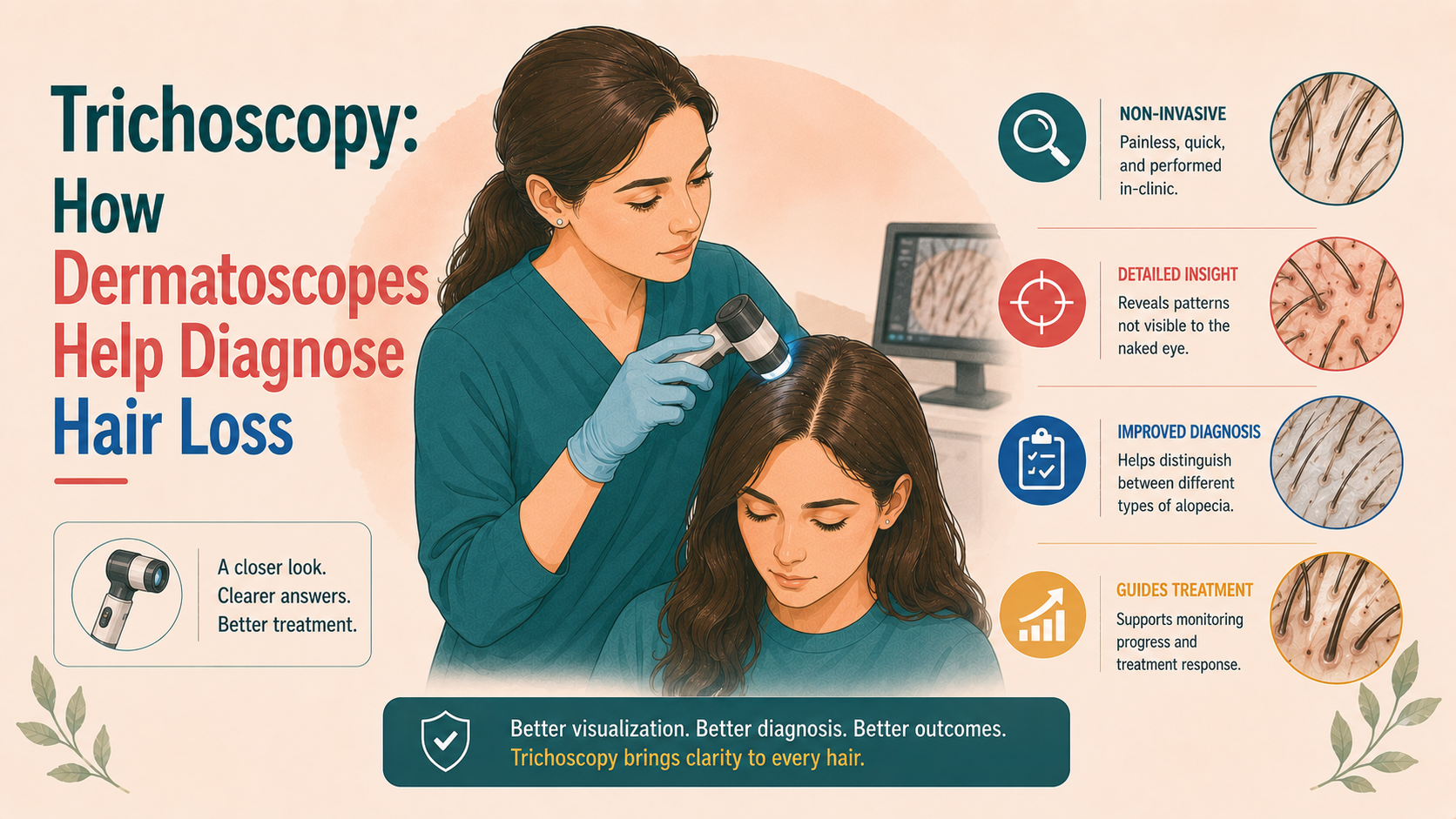
Trichoscopy is the use of a handheld or video dermatoscope to examine the scalp and hair at magnifications typically between 10× and 70×. It can be performed using contact or non-contact techniques, polarised or non-polarised light, and is increasingly integrated with digital image capture and software-based analysis.
At these magnifications, the clinician can visualise hair shafts in cross-section and along their length, follicular openings (ostia) and perifollicular structures, as well as small blood vessels and pigmentation patterns in the scalp skin.
Before trichoscopy, decisions about biopsy and treatment often relied on clinical pattern alone. Trichoscopy has allowed:
- earlier distinction between scarring and non-scarring alopecias;
- recognition of characteristic “signatures” in different conditions;
- more targeted biopsies when needed;
- and more objective monitoring of response to therapy.
It has become an extension of the clinical examination rather than an optional extra.
2. How trichoscopy is done in practice
In the clinic, trichoscopy typically looks like this:
- the doctor places a dermatoscope against the scalp (sometimes with a drop of alcohol or gel for better contact) or holds it just above the surface;
- they move systematically across regions: frontal, temporal, vertex, occipital, and any areas of focal concern;
- images may be captured for the record and for later comparison.
The technique is quick and non-invasive. No hair needs to be plucked, and for most patients, it is entirely painless. It is best thought of as a magnifying window into what the hair and scalp are doing at a microscopic level.
3. Key trichoscopic structures
On trichoscopy, the clinician pays attention to several recurring elements:
- Hair shaft diameter and shape – whether shafts are of uniform thickness or mixed, and whether there are bends, twists, nodes or breakages.
- Follicular openings (ostia) – whether they are present and filled with one or more hairs, or absent (suggesting scarring).
- Perifollicular features – such as pigmentation (“peripilar sign”), white halos, scale or “collarettes”.
- Background scalp skin – colour, pigment network, vascular patterns.
- Empty follicles and regrowing hairs – appearance of newly emerging thin hairs, often labelled “upright regrowing hairs”.
Different diseases produce different combinations of these patterns. Recognising them is what gives trichoscopy its diagnostic power.
4. Androgenetic alopecia and female pattern hair loss
Androgenetic alopecia (AGA) in men and female pattern hair loss (FPHL) share trichoscopic hallmarks.
The most characteristic features are:
- Hair shaft diameter diversity – in affected areas, hairs vary widely in thickness, with a mix of normal terminal hairs, thinner intermediate hairs and very fine vellus-like hairs. This heterogeneity, usually >20% variation in diameter, reflects miniaturisation.
- Increased proportion of vellus-like hairs – short, thin, lightly pigmented shafts in the same field as normal hairs.
- Yellow dots – follicular openings filled with sebum and keratin, appearing as small, round, yellowish structures; more prominent in advanced disease.
- Peripilar sign – a brownish halo or subtle pigmentation around the proximal shaft, thought to correspond to perifollicular inflammation and melanin incontinence.
- Empty-looking follicles – in more advanced AGA, areas with reduced numbers of visible hairs per ostium.
In men, these changes are most pronounced in the frontal and vertex areas, with the occipital region serving as an internal control (more uniform shafts, fewer vellus hairs). In women, they are concentrated along the central part and crown, with relative sparing of the lower occipital scalp.
Trichoscopy in AGA/FPHL is especially helpful in:
- early disease, when naked-eye changes are subtle;
- distinguishing pattern hair loss from diffuse telogen effluvium and alopecia areata incognita;
- tracking miniaturisation and response to therapy over time.
5. Telogen effluvium
Telogen effluvium (TE) is a reactive shedding rather than a primary miniaturising process. On trichoscopy:
- Hair shaft diameter is relatively uniform – there is not the pronounced heterogeneity seen in AGA.
- Follicular openings are preserved, with roughly equal numbers of hairs per ostium compared to controls; in chronic TE, there may be more empty ostia but without the marked vellus proliferation of AGA.
- Increased number of short regrowing hairs (“upright regrowing hairs”) can be seen as TE resolves.
- Background scalp features are usually normal.
Clinically, TE and early FPHL can look similar in women with diffuse thinning. Here, trichoscopy contributes by highlighting the presence or absence of miniaturisation and guiding whether to prioritise trigger identification and supportive measures (as in TE) or anti-androgen/minoxidil strategies (as in FPHL).
6. Alopecia areata
Alopecia areata (AA) has some of the most recognisable trichoscopic signatures.
In patchy AA, trichoscopy may show:
- Yellow dots – numerous, dense, often uniform; in AA, they represent empty follicles filled with degenerating keratin and sebum.
- Black dots – pigmented residues of broken hairs at the follicular opening.
- Exclamation mark hairs – short hairs tapered proximally (narrower at the scalp), reflecting proximal shaft weakness.
- Broken hairs of various lengths.
- Tapered hairs emerging at the periphery of patches.
In diffuse forms (alopecia areata incognita), patterns can resemble TE, but the presence of numerous yellow dots and black dots, even in the absence of clear patches, points towards AA.
These findings are useful for:
- differentiating AA from trichotillomania (where more varied broken hair patterns and specific “flame” or “tulip” hairs may be seen);
- assessing disease activity (presence of exclamation mark hairs and black dots suggests ongoing activity);
- guiding biopsy when needed.
7. Traction alopecia and trichotillomania
7.1 Traction alopecia
Traction alopecia typically affects the frontal and temporal hairline in individuals wearing tight hairstyles. Trichoscopy can reveal:
- Reduced hair density in affected areas;
- Hair of varying lengths, including short, broken hairs;
- Early on, preserved follicular openings; in chronic cases, loss of some ostia and signs of scarring;
- Sometimes “fringe sign” – retention of thin vellus hairs along the hairline.
The combination of clinical history and trichoscopy helps distinguish traction from frontal fibrosing alopecia and other scarring conditions.
7.2 Trichotillomania
Trichotillomania, a hair-pulling disorder, has a distinct trichoscopic pattern:
- Broken hairs at different lengths – often with irregular fracture points;
- Coiled hairs and “hook hairs” where shafts are bent by mechanical trauma;
- Flame hairs – semi-transparent, cone-shaped residues;
- Tulip hairs – dark-ended, tapered hairs resembling a tulip;
- Empty follicles and haemorrhagic spots in some areas.
There is typically marked variability within a small field – normal hairs next to a variety of broken and distorted shafts. This asymmetry helps differentiate trichotillomania from AA and TE.
8. Tinea capitis and infectious processes
In tinea capitis (scalp dermatophyte infection), especially in children, trichoscopy can show:
- Comma hairs – curved, comma-shaped, short hairs;
- Corkscrew hairs – spiral-shaped hairs, often seen in African hair types;
- Morse-code hairs – alternating light and dark segments along the shaft;
- Black dots – broken-off hairs at the scalp surface.
This pattern is an important adjunct, particularly when scaling and lymphadenopathy (swollen glands) are subtle. It supports early diagnosis and treatment without waiting for culture.
In folliculitis decalvans and dissecting cellulitis, trichoscopy may reveal clusters of hair (“tufts”) emerging from inflamed or scarred follicles, pustules, and the absence of openings in advanced areas.
9. Scarring (cicatricial) alopecias
Scarring alopecias involve permanent destruction of the follicle and replacement with fibrous tissue. Trichoscopy is particularly helpful in recognising early scarring before naked-eye atrophy is obvious.
General scarring features include:
- Loss of follicular openings – appearing as white patches or structureless areas;
- Perifollicular scale and erythema around remaining hairs;
- Perifollicular white halos (“peripilar casts”);
- Blue-grey dots in some lichenoid conditions, reflecting pigment incontinence.
Specific conditions have their nuances:
9.1 Lichen planopilaris / frontal fibrosing alopecia
- Perifollicular whitish scale (“perifollicular casts”) at the base of hairs;
- Prominent perifollicular erythema;
- Appearance of isolated, lonely hairs in areas otherwise devoid of follicular openings;
- In FFA, involvement of the frontal hairline and eyebrows.
9.2 Central centrifugal cicatricial alopecia (CCCA)
- Perifollicular white or grey scale at the central scalp;
- Reduced follicular density radiating outwards;
- “Peripilar halos” and increased interfollicular white patches in more advanced disease.
9.3 Discoid lupus erythematosus
- Thick adherent scale plugging follicular openings;
- Arborising (branching) vessels;
- White patches and follicular dropout;
- Pigmentary changes, including blue-grey dots at the periphery.
Recognising these patterns allows earlier suspicion of scarring disease, targeted biopsy of the most informative area, and timely initiation of immunomodulatory therapy to prevent further permanent loss.
10. Quantitative trichoscopy and phototrichograms
Beyond pattern recognition, trichoscopy can be used quantitatively:
- Phototrichogram techniques involve clipping a small area, photographing it at baseline and after a set interval, and counting hairs to assess growth (anagen vs telogen).
- Software can analyse stored trichoscopic images to estimate hair density, shaft diameter distribution and miniaturisation index.
These methods are useful in clinical trials, tracking responses to therapies such as finasteride, minoxidil, and PRP, and providing more objective documentation in private practice.
For patients, this means that treatment response can sometimes be quantified using numbers and images, rather than solely subjective impressions.
11. Limitations and appropriate use
Trichoscopy is a powerful tool, but not infallible. Limitations include:
- learning curve – interpretation requires training and experience;
- overlapping features between conditions in some cases;
- optical artefacts from gel, hair products or lighting;
- lack of standardisation in some quantitative methods.
However, it does not replace a thorough history and physical examination, appropriate laboratory testing, or histopathological confirmation in cases of diagnostic uncertainty or scarring. Rather, it acts as a bridge between what the naked eye sees and what the biopsy reveals.
12. What this means for patients
If your dermatologist uses a dermatoscope to examine your scalp, they are:
- looking for specific patterns that can narrow the diagnosis;
- deciding whether biopsy is necessary and, if so, where to perform it;
- establishing a visual baseline for future comparisons.
You can reasonably expect that in a specialist hair clinic, trichoscopy will be part of standard assessment. If you have a complex or unclear case and trichoscopy is not mentioned, it is appropriate to ask whether it could provide useful information.

![[headshot]](https://cdn.prod.website-files.com/68207da82e5b8c350c67932f/68c6a7436a552c0fe87a4da7_Screenshot%202024-11-27%20at%2011.38.11.png)