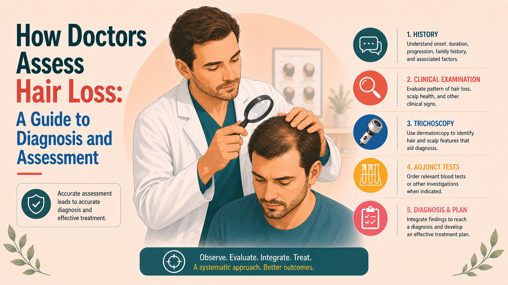
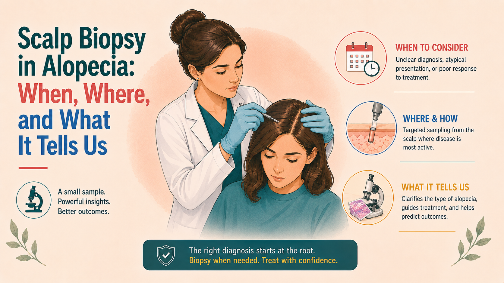
A small sample of scalp skin examined under a microscope to clarify uncertain hair loss diagnoses.
A scalp biopsy is not needed for every hair loss patient, but it can be decisive when the diagnosis is uncertain or scarring alopecia is suspected. The sample is usually taken from an active edge rather than a completely bald scarred area. Histology can distinguish non-scarring from scarring alopecia, show inflammation pattern, and support diagnoses such as lichen planopilaris, frontal fibrosing alopecia or diffuse alopecia areata. In pattern hair loss, biopsy may show miniaturised follicles and altered terminal-to-vellus ratios. The result is only as useful as the chosen biopsy site.
Starace M et al. Female Androgenetic Alopecia: Diagnosis and Management, Am J Clin Dermatol, 2020 — https://doi.org/10.1007/s40257-019-00479-x; Anastassakis K. Androgenetic Alopecia From A to Z, Vol. 1, Springer, 2022 — https://doi.org/10.1007/978-3-030-76111-0; British Association of Dermatologists: Hair loss female pattern (androgenetic alopecia) — https://www.bad.org.uk/pils/hair-loss-female-pattern-androgenetic-alopecia